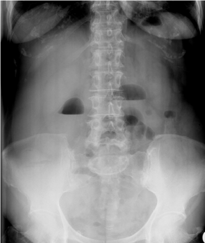
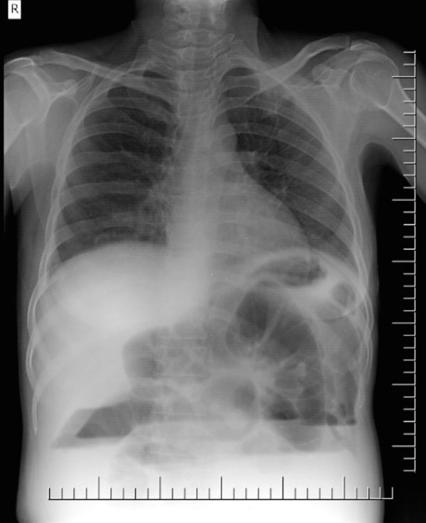
Lung Bullae With AirFluid Levels What Is the Appropriate Therapeutic Approach? Respiratory Care

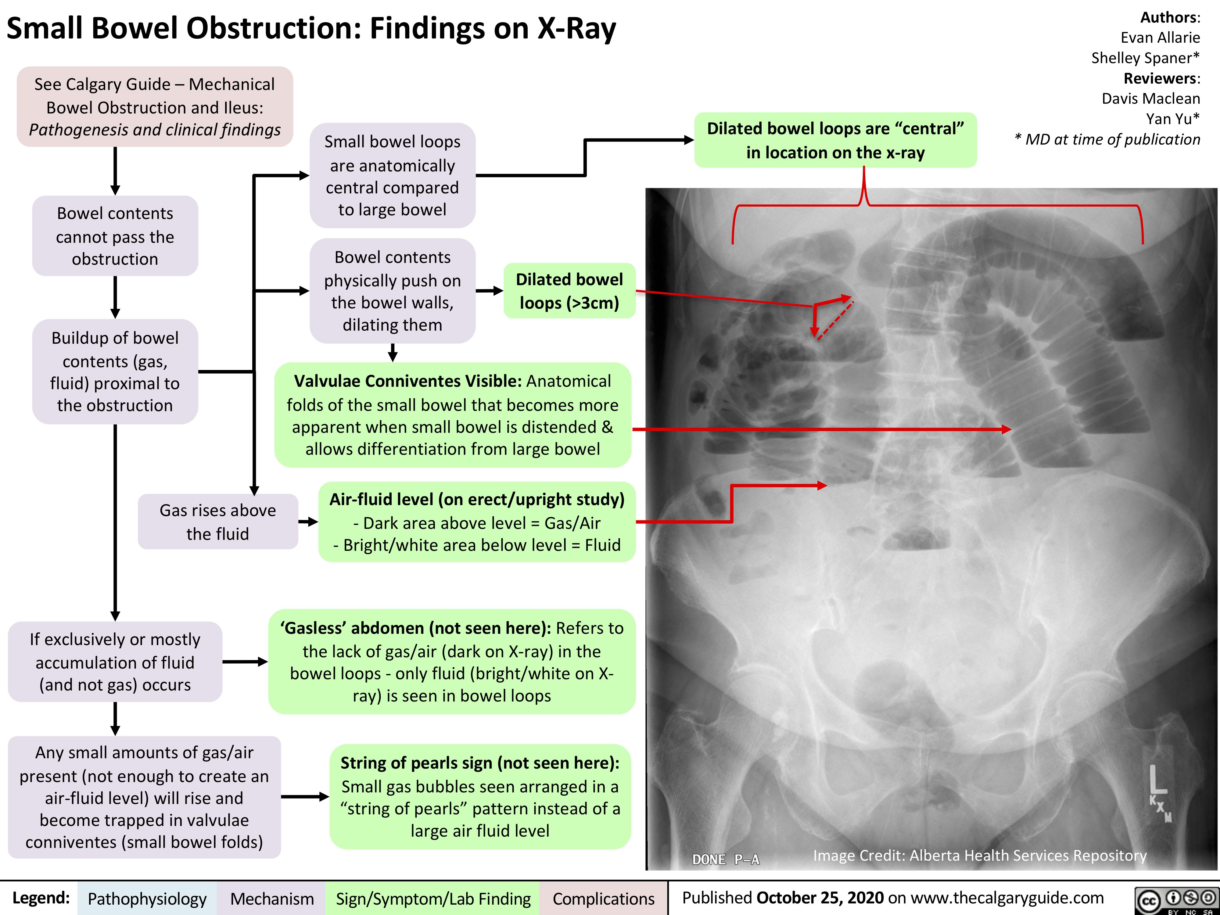
Small Bowel Obstruction Air Fluid Levels
Pendekatan diagnosis harus mempertimbangkan gambaran klinis, karakteristik pasien, dan gambaran kavitas pada pemeriksaan radiologi. [1] Penyebab Infeksi. Dalam tahap awal evaluasi lesi kavitas rontgen toraks, penting untuk menentukan terlebih dulu apakah lesi disebabkan oleh penyebab infeksius atau tidak.

Bronchial atresia a rare presentation as air fluid level on chest roentgenogram BMJ Case Reports
ditemukan gambaran air fluid level pad a. penderita sinusitis maksilaris. Pada pen elitian. oleh W ardani et al (2 014) 7 orang penderita (15,56 %) dengan penebalan mukosa, 15 orang.

Chest Radiography Xray Show Beautiful Airfluid Stock Photo 1357114544 Shutterstock
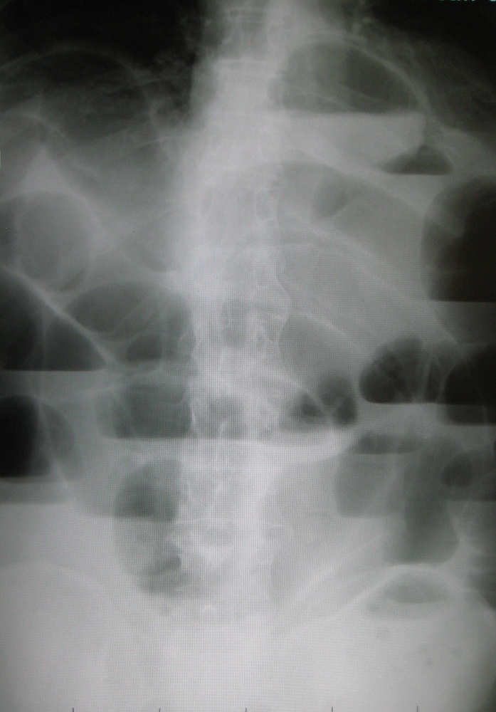
An air-fluid level in bowel on X-ray is a common finding that can represent a normal finding all the way to life threatening bowel obstruction. An air-fluid level means that there is both air and fluid in a bowel loop. The air will rise to the top of the bowel loop and the fluid will settle at the bottom. There is a sharp straight interface.

Airfluid levels in abdominal Xray graphy. Download Scientific Diagram
Bronchiectasis is the common response of bronchi to a combination of inflammation and obstruction/impaired clearance. Causes include 1-7,9,17,21: idiopathic (most common) impaired host defenses. cystic fibrosis (most common cause in children) primary ciliary dyskinesia, e.g. Kartagener syndrome , Young syndrome.

Chest xray on admission showing an unusual air fluid level in the... Download Scientific Diagram
Air fluid level Efusi pleura Daerah avaskuler; Air fluid level; Fluidopneumothorax Gambaran sarang tawon (honeycomb appearance) di hemithorax dextra; hemithorax sinistra; Bronkhiektasis dextra; sinistra; Gambaran sayap kelelawar/kupu-kupu (bat/butterfly's wing appearance) Uremic lung

Lateral plain Xray showing airfluid level in the maxillary sinus. Download Scientific Diagram
gasless abdomen: gas within the small bowel is a function of vomiting, NG tube placement and level of obstruction. string-of-beads sign: small pockets of gas within a fluid-filled small bowel. Ultrasound. Bedside tests help to diagnose small bowel obstruction, findings suggestive of small bowel obstruction 7: dilated bowel loop (diameter > 3 cm)

Small Bowel Obstruction Air Fluid Levels
Citation, DOI, disclosures and article data. Stepladder sign represents the appearance of distended small bowel loops with gas-fluid levels that appear to be stacked on top of each other, typically observed on erect abdominal radiographs in the setting of small bowel obstruction .

AIR FLUID LEVELS
The conventional paranasal sinus examination should consist of a minimum of three views: the Caldwell (posteroanterior), Waters (occipitomental), and lateral views (Figs. 36-1, 36-2, and 36-3).The primary purpose of the Caldwell view is to visualize the frontal and ethmoid sinuses, whereas the maxillary sinuses are best demonstrated with the Waters view.

Air Fluid Level in Gastric Distention on X Ray YouTube
Mengingat abses paru merupakan salah satu bentuk infeksi pada paru-paru, diagnosis banding yang perlu dipikirkan adalah infeksi lain seperti tuberkulosis. Selain itu, diagnosis banding lainnya yang perlu dipertimbangkan adalah karsinoma berkavitas, granulomatosis Wegener, kista atau bulla terinfeksi, dan aspergilloma. [2,6,18] Tuberkulosis.

Lung Bullae With AirFluid Levels What Is the Appropriate Therapeutic Approach? Respiratory Care
Bullous emphysema is characterized by the presence of air-filled lung parenchymal spaces termed 'emphysematous bullae' which result from the destruction of the alveolar spaces. Rarely, they may be present as isolated spaces in otherwise normal lungs. [ 2] Drouet et al. first described the air-fluid level in emphysematous bullae in 1947 as.

X ray of the abdomen showed multiple air fluid levels without any gas... Download Scientific
Importance of differential air-fluid levels on plain radiographs in the diagnosis of bowel obstruction. Author : D Bryk Author Info & Affiliations Volume 163 , Issue 1

Abdominal XRay with Air Fluid Levels REBEL EM Emergency Medicine Blog
Gambaran air fluid level pada cavitas menunjukkan kemungkinan adanya superinfeksi di dalam cavitas.. (fungus ball) atau mycetoma yang memberikan gambaran air crescent sign pada foto thorax dan dapat juga memenuhi seluruh cavitas. Efusi pleura (komplikasi) Pada TB Primer: unilateral, efusi pleura luas tanpa lokulasi.

x ray air fluid level
Posisi terlentang (supine) Pelebaran usus di proksimal daerah obstruksi Penebalan dinding usus, Herring Bone Appearance (duri ikan). Gambaran ini didapat dari pengumpulan gas dalam lumen usus yang melebar 2. Posisi setengah duduk atau berdiri. Air fluid level Step ladder appearance. 3.

PPT Diagnostic Radiology III Definitions PowerPoint Presentation ID411346
Gambaran CT pada abses paru adalah kavitas yang terlihat bulat dengan dinding tebal, tidak teratur, terletak di daerah jaringan paru yang rusak dan tampak gambaran air-fluid level. Tampak bronkus dan pembuluh darah paru berakhir secara mendadak pada dinding abses, tidak tertekan atau berpindah letak.

Air Fluid Level PDF
An air bronchogram occurs when endobronchial air is visible against a background of increased lung opacity. Expulsion of gas from the parenchyma is partial or complete and can be due to atelectasis and/or replacement by fluid, inflammatory cells, blood, tumor or interstitial thickening. The persistence of gas in the bronchi implies patency of.

x ray air fluid level
memberikan gambaran bulat dengan radioluscent tanpa corakan paru. Kadang kavitas dapat berisi cairan yang merupakan produk radang yang memberikan gambaran air fluid level.14 Kavitas jarang ditemukan karena berdasarkan patofisiologi terjadinya tuberkulosis paru, jika sudah terjadi fokus primer yaitu dimana kuman